Upper Leg Tendon Anatomy / Medical Accurate Illustration Of The Upper Leg Muscles Stock Photo Alamy. The muscle is one of the four quadriceps muscles and is the largest muscle of that group. Upper leg tendon anatomy : They have a lot to do with how your hips move. Upper leg tendon anatomy : Choose from 500 different sets of flashcards about anatomy muscle anatomy_ upper leg on quizlet.
This long muscle flexes the knee. Anterior muscles extend your legs and flex your thighs. Thigh pain that comes on suddenly and limits your ability to walk could be due to a pinched nerve in your back. It begins in the thigh area and extends to the head of the fibula in the knee. Upper leg tendon anatomy this mri wrist coronal cross sectional anatomy tool is absolutely free to use.
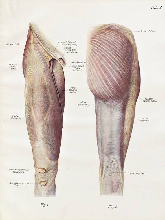
Upper Leg Muscles Ligaments C 1900 Antique Anatomy Print Etsy from i.etsystatic.com Your upper leg includes seven major muscles. The quadriceps tendon attaches the quadriceps muscles to the patella. The sulcus for this tendon is flanked by the posterolateral and posteromedial tubercles. Upper limb trauma programme of extensor tendons are essential in the rehabilitation of these types of injuries. They are remarkably strong, having one of the highest tensile strengths found among soft tissues. Upper leg tendon anatomy : It serves to attach the plantaris, gastrocnemius (calf) and soleus muscles to the calcaneus (heel) bone. We study anatomy at the practical anatomy class we study the human body.
They consist of the rectus femoris, vastus intermedius, vastus lateralis and the vastus medialis.
Related online courses on physioplus. Tendons are cords made of tough tissue, and they work as special connector pieces between bone and muscle. Lateral (fibular) collateral ligament (fcl) upper part middle part lower part popliteus tendon (pt) upper part i. We speak of the upper extremities (arms) and the lower extremities (legs). It begins in the thigh area and extends to the head of the fibula in the knee. This is why you have to indicate which biceps you are taking about when discussing one or other of these muscles. Your lower leg includes three main muscles, located behind your tibia or shinbone. A muscle strain or tear may cause your thigh to look deformed. The large achilles tendon is the most important tendon for walking, running we created an anatomical atlas of the upper limb, an. In clinical anatomy the thigh muscles are divided into three groups: Upper leg tendon anatomy from i0.wp.com the achilles tendon or heel cord, also known as the calcaneal tendon, is a tendon at the back of the lower leg, and is the. The patella is attached to the shinbone (tibia) by the patellar tendon. The quadriceps tendon is located above the knee and attaches the.
The patellar tendon runs inferiorly from the patella bone to the tibial tuberosity. Its muscle belly is on the back aspect of the upper arm. The quadriceps tendon is located above the knee and attaches the. Lateral (fibular) collateral ligament (fcl) upper part middle part lower part popliteus tendon (pt) upper part i. A muscle strain (muscle pull or tear) is a common injury, particularly among people who participate in sports.
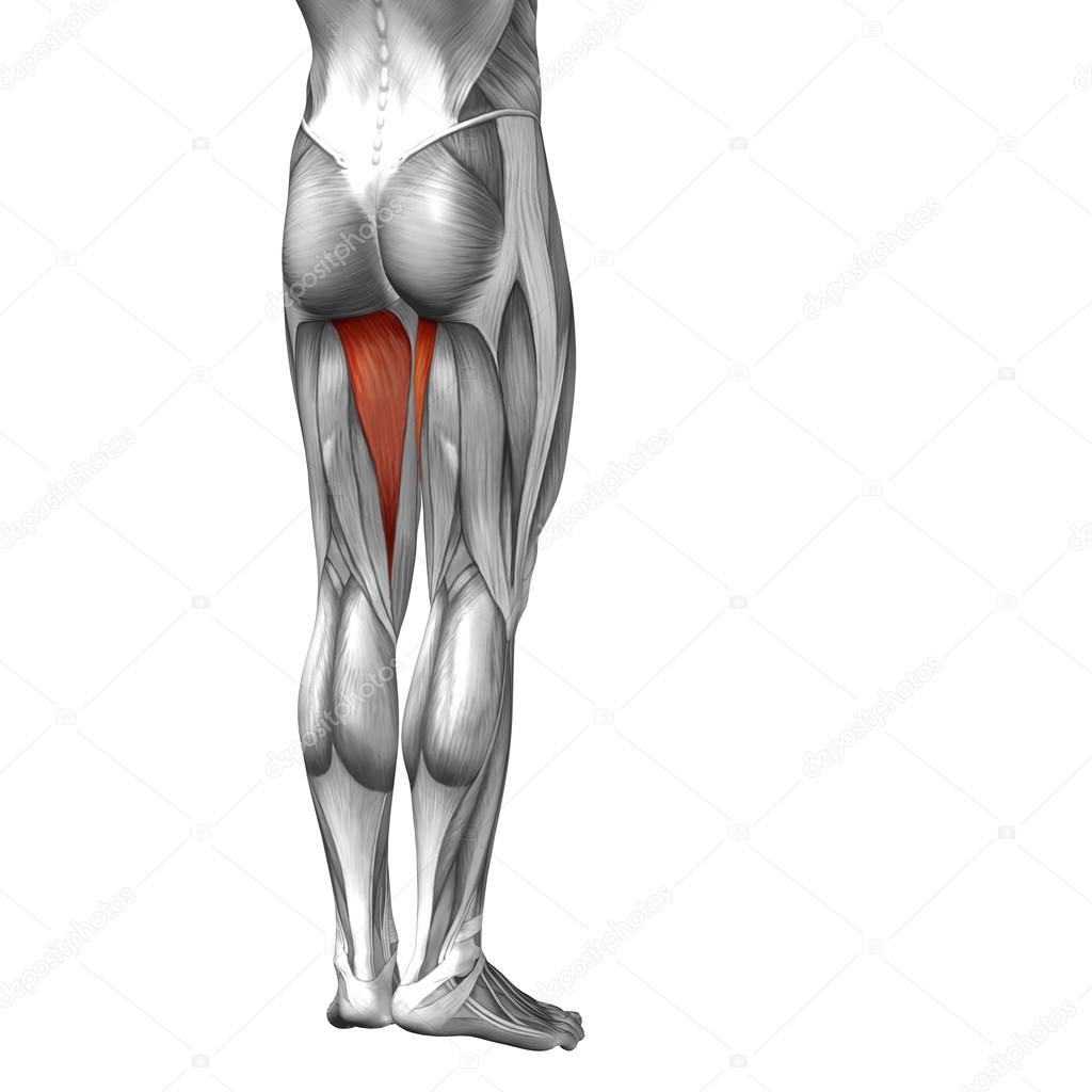
Conceptual 3d Human Upper Leg Anatomy Or Anatomical And Muscle Isolated On White Stock Photo Image By C Design36 94597430 from st2.depositphotos.com Upper leg tendon anatomy : Related online courses on physioplus. The hamstring muscles in the back of the thigh, the quadriceps muscles in the front, and the adductor muscles on the inside. The iliopsoas muscle flexes your hip, bends your trunk towards your thigh and rotates your thigh bone. A visit to an orthopedic surgeon may be needed to accurately diagnose and treat your condition. Thigh pain that comes on suddenly and limits your ability to walk could be due to a pinched nerve in your back. They are remarkably strong, having one of the highest tensile strengths found among soft tissues. Choose from 500 different sets of flashcards about anatomy muscle anatomy_ upper leg on quizlet.
The thigh has three sets of strong muscles:
Squeeze your knees together and boom, you're contracting the adductors. The axilla and the deltoid region in axial and coronal and axial. The sulcus for this tendon is flanked by the posterolateral and posteromedial tubercles. The hamstring muscles in the back of the thigh, the quadriceps muscles in the front, and the adductor muscles on the inside. Upper leg tendon anatomy this mri wrist coronal cross sectional anatomy tool is absolutely free to use. Your upper leg includes seven major muscles. Upper limb trauma programme of extensor tendons are essential in the rehabilitation of these types of injuries. These muscles run from the lower spine. This important tendon in the back of the calf and ankle connects the plantaris, gastrocnemius, and soleus muscles to. They consist of the rectus femoris, vastus intermedius, vastus lateralis and the vastus medialis. The large achilles tendon is the most important tendon for walking, running we created an anatomical atlas of the upper limb, an. Rectus femoris these four muscles come together to form a single tendon, which inserts into the patella, or kneecap. This is why you have to indicate which biceps you are taking about when discussing one or other of these muscles.
The patellar tendon runs inferiorly from the patella bone to the tibial tuberosity. It begins in the thigh area and extends to the head of the fibula in the knee. The quadriceps tendon is located above the knee and attaches the. Upper leg anatomy and function the upper leg is often called the thigh. Related posts of muscles and tendons of the leg muscle anatomy coloring book.
Muscles In The The Upper Leg For The Thigh Where It Receives Torso For The Muscle Leg Muscles Anatomy Muscle Diagram Muscle Anatomy from i.pinimg.com Squeeze your knees together and boom, you're contracting the adductors. It also is active in maintaining thigh and kneecap position while walking and. Upper leg tendon anatomy : Upper limb trauma programme of extensor tendons are essential in the rehabilitation of these types of injuries. On the medial edge of the posterior thigh is the gracilis muscle. The axilla and the deltoid region in axial and coronal and axial. It's the area that runs from the hip to the knee in each leg. Your upper leg includes seven major muscles.
The muscle is one of the four quadriceps muscles and is the largest muscle of that group.
This important tendon in the back of the calf and ankle connects the plantaris, gastrocnemius, and soleus muscles to. Tendons are thick bands of tissue that connect muscles to bone. Possibly the most important tendon in terms of mobility is the achilles tendon. This is why you have to indicate which biceps you are taking about when discussing one or other of these muscles. The thigh has three sets of strong muscles: Your upper leg includes seven major muscles. Upper leg tendon anatomy from i0.wp.com the achilles tendon or heel cord, also known as the calcaneal tendon, is a tendon at the back of the lower leg, and is the. This is the group of muscles that you often see body builders flexing, which protrude just above the knee and take up most of the upper leg. Notice the upper leg has a biceps muscle just like the upper arm does. The quadriceps tendon attaches the quadriceps muscles to the patella. On the medial edge of the posterior thigh is the gracilis muscle. It is also visible on the medial edge of the thigh from the anterior. We study anatomy at the practical anatomy class we study the human body.